What you’re getting yourself into
6,300 words, 21-42 minute read time
Key Points
- Sarcoplasmic hypertrophy – growth of the sarcoplasm that outpaces the growth of the myofibrils – does seem to happen to a significant degree.
- Simple increases in glycogen storage don’t seem to be the primary driver of sarcoplasmic hypertrophy. It seems to be driven by an increase in sarcoplasmic protein content.
- The degree to which sarcoplasmic hypertrophy takes place may be influenced by training, but whether you can specifically train for sarcoplasmic vs. myofibrillar hypertrophy is unclear. More than anything, it seems to be a natural consequence of muscle growth itself.
- Looking at strength differences between bodybuilders and powerlifters or weightlifters is not a valid way to assess degree of sarcoplasmic hypertrophy. However, dismissing sarcoplasmic hypertrophy simply because there are better explanations for the observed differences in relative strength is foolhardy.
- It’s unclear whether steroids increase the amount of sarcoplasmic hypertrophy that takes place.
You may have heard the old piece of bodybuilding forum “wisdom”: “Bodybuilders are bigger than powerlifters/weightlifters but still lift less because they have more sarcoplasmic hypertrophy, bro.”
You may have seen pictures like this before as well, from very credible sources:
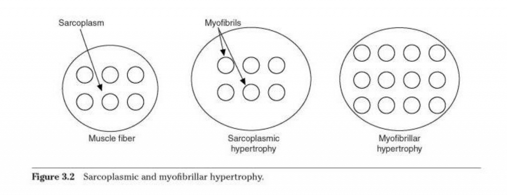

If you’re not sure what we’re even talking about here, let me catch you up:
It’s been proposed that there are two ways your muscle fibers can grow:
- Myofibrillar hypertrophy, which is accomplished via the growth and multiplication of the myofibrils inside each muscle fiber. The myofibrils are the actual “motors” of the muscle fiber, made up of contractile proteins that make the muscle fiber contract. This is illustrated by the picture on the far right in the image above.
- Sarcoplasmic hypertrophy, which is, in theory, accomplished by the expansion of the sarcoplasm (the cytoplasm of the muscle) inside the muscle fiber. This is illustrated by the middle picture in the image above.
Now, just to define these terms as accurately as possible, no one is suggesting that the sarcoplasm can’t expand at all. Rather, myofibrillar hypertrophy implies that the sarcoplasm expands at roughly the same rate as the myofibrils grow and expand (so if myofibrils took up 80% of the room within the muscle fiber before, and the sarcoplasm took up 20% of the room within the muscle fiber, with a doubling in fiber size, that proportion would still hold), and sarcoplasmic hypertrophy implies that the sarcoplasm expands at a significantly faster rate than the myofibrils grow and divide (so if there was an 80/20 ratio initially, it may be 70/30 or 60/40 after sarcoplasmic hypertrophy).
So, does sarcoplasmic hypertrophy happen, and does it meaningfully contribute to overall muscle growth? That’s the million dollar question.
I’ve been skeptical of sarcoplasmic hypertrophy for a long time, mainly because it wasn’t an adequate explanation for the problem it sought to address.
The situation where sarcoplasmic hypertrophy is most often invoked is when comparing bodybuilders to powerlifters or weightlifters. How can some 280 lb. bodybuilder get out-squatted by a 180 lb. strength athlete? Sarcoplasmic hypertrophy explains this difference, the thinking goes. The bodybuilders must just have “non-functional” sarcoplasmic hypertrophy, making their muscles bigger without making them stronger since it’s the myofibrils that contain the contractile proteins.
However, as I’m sure any consistent reader of this site is aware, strength also has a massive skill component. The strength athletes are consistently squatting really heavy loads in training, so they’ll generally be more skillful squatters. If the bodybuilders changed their training style for a few months, their squat numbers would skyrocket. You can see this both in the real world (it didn’t take too long for Stan Efferding to squat 900+ after switching from bodybuilding to powerlifting, for example), and in scientific research (it’s a consistent enough finding that strength is pattern-specific and load-specific that it’s not even worth citing specific studies).
On second reflection, though, I realized that I may have been in the wrong for dismissing sarcoplasmic hypertrophy just because it’s a poor explanation for the phenomenon it’s invoked to explain. Expressed more formally, if someone claims “A causes C,” but B is actually a more likely cause for C than A, you can’t then conclude that A doesn’t exist. You just conclude that B is a better explanation for C than A is.
Here’s a ridiculous example to make this point clear: If someone claimed that “cats caused that building to explode,” but it was later discovered that there was a leak in the gas main that ignited, you could only conclude that the gas catching fire was a better explanation for the explosion. You could not conclude that cats don’t exist.
So, the main argument for sarcoplasmic hypertrophy falls flat (because there’s a better explanation for the strength difference), and the main argument against it is based on somewhat sneaky logical fallacy.
With all of that out of the way, we can start to really dig into this topic with a clean slate.
So, first of all, when people talk about sarcoplasmic hypertrophy, they’re talking about something that’s long-lasting, directly influenced by training, and that makes a meaningful difference in muscle size. In other words, you can get some increases in sarcoplasmic volume by taking creatine, carb loading, by doing blood flow restriction training, or by causing muscle damage, but none of these things cause the type of sarcoplasmic hypertrophy people are interested in. None of them cause a very large increase in muscle volume in the first place, and they’re all transient. Stop taking creatine or carb loading, and the water goes away. The fluid retention in the muscles after a session of blood flow restriction training or a training session that causes a lot muscle damage dissipates within 72 hours, and the effect gets progressively smaller over time.
We first have to ask ourselves: Where could this sarcoplasmic hypertrophy be coming from in the first place?
There are two primary places:
- An increase in osmotic non-protein solutes in the muscle fiber, which will then attract more water.
- An increase of sarcoplasmic proteins (including all organelles except for nuclei and myofibrils) relative to contractile proteins.
Solutes in the Muscle
Increasing solutes inside the muscle is the first way that you could potentially induce sarcoplasmic hypertrophy.
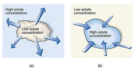
I’m sure you remember pictures like this from high school biology:
If you could cause sarcoplasmic hypertrophy by increasing the amount of solutes inside the muscle fibers, it would be a scenario like the picture on the right. Since water follows solutes, and since muscle fibers are permeable to water, if you added more “stuff” (other than contractile proteins) inside the muscle fiber, water would follow, and voila, sarcoplasmic hypertrophy.
Unfortunately, that’s really not feasible for non-protein solutes.
Ion concentrations (sodium, potassium, bicarbonate, calcium, hydrogen, etc.) won’t change too much long-term as long as you have healthy kidneys.
You have some fatty acids stored in muscle fibers as well, and those lipid droplets can grow and divide with training (specifically aerobic training), but they don’t attract much water, and they make up such a small fraction of the space inside a muscle fiber that there’s really no way they could make much of a difference.
Finally, you have glycogen. And yes, glycogen storage capacity can increase with training. However, maximal glycogen concentrations can increase with just about any type of training; they may just increase a bit more with aerobic training or the type of lifting people think will cause sarcoplasmic hypertrophy (lighter, high volume, bodybuilder-style training). Ultimately, glycogen concentrations simply can’t make a huge difference either, though. Even when you infuse glucose and insulin directly into a muscle for 8 hours, glycogen concentrations peak at about 4 grams of glycogen per 100 grams of muscle. One gram of glycogen is stored with 3 grams of water, so, at most, muscle glycogen plus the water associated with it makes up 16% of the total mass of a muscle.
Average skeletal muscle glycogen concentrations are closer to 1.5-2 grams of glycogen per 100 grams of muscle. In other words, going from “average” glycogen concentrations to 100% maxed-out glycogen concentrations would increase total muscle mass by about 6-8%, and that increase would be “sarcoplasmic hypertrophy.” However, a) a 6-8% increase isn’t a huge increase (certainly not as large of an increase as most people expect), and b) since regular strength training increases glycogen storage capacity in the first place, the difference in maximum glycogen concentrations would make a difference of maybe 2-3%, not 6-8%. Ultimately, glycogen concentrations are more influenced by diet than by training, and, again, any increase would be transient, so this doesn’t fit the bill.*
*Note that glycogen doesn’t fit the bill because its effects are transient. If you go from fully depleted to fully repleted, that can make a noticeable difference (compare pictures of bodybuilders in their last few weeks of dieting versus how they look on stage for a dramatic illustration), but it’s not a permanent change, and training style doesn’t affect how much of a glycogen pump you can get to a tremendous degree.
Non-Contractile Proteins
Could an increase in non-contractile proteins relative to contractile proteins cause sarcoplasmic hypertrophy?
Maybe.
Protein and glycogen draw similar amounts of water into muscle fibers (1 gram of each is stored with about 3 grams of water). So, if the concentration of sarcoplasmic proteins is higher than the concentration of glycogen (so that they could make a more meaningful difference in overall muscle size), and the concentration of sarcoplasmic proteins can be altered with training, maybe that would be a reasonable avenue by which sarcoplasmic hypertrophy could take place.
It was surprisingly difficult to find a source that compared the total amount of sarcoplasmic and myofibrillar proteins in skeletal muscle. I’m guessing that most of that work is really old, and really buried in the PubMed and Google Scholar search results. An introductory textbook about meat science (i.e. for people who want to work in the meat processing industry) said that concentrations of myofibrillar proteins were about 3x higher than concentrations of sarcoplasmic proteins in mammals. That roughly matches a study on guinea pigs and rabbits that was published way back in the ’60s.
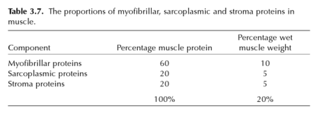
Now we’re getting somewhere. If a 3:1 ratio is typical, then maybe that spread could change.
Most studies that measure rates of myofibrillar and sarcoplasmic protein synthesis separately find that they don’t always follow the exact same pattern in response to the same stimulus. However, most of this data actually weakens the case for sarcoplasmic hypertrophy. Although lifting increases protein synthesis rates for both myofibrillar and sarcoplasmic proteins, the increase is generally larger and more sustained for myofibrillar proteins. The only two major instances where sarcoplasmic protein balance consistently has the advantage are inactivity and aging; sarcoplasmic protein breakdown proceeds at a slower rate with complete unloading, and sarcoplasmic protein synthesis doesn’t decrease with age, whereas myofibrillar protein synthesis does. Also, we need to keep in mind that measuring rates of protein synthesis doesn’t necessarily tell us much about long-term hypertrophy.
However, these studies do show that sarcoplasmic protein concentrations in the muscle aren’t pegged to myofibrillar protein concentrations in any major way. In fact, in the rabbit study referenced earlier, the researchers saw a pretty big spread in the ratio of myofibrillar and sarcoplasmic proteins. In young, active rabbits, the ratio was a bit under the standard 3:1 – about 2.4:1. However, in rabbits that were forced to be inactive, that ratio shifted to about 1.6:1, and by middle age, the rabbits actually had slightly higher concentrations of sarcoplasmic proteins than myofibrillar proteins.
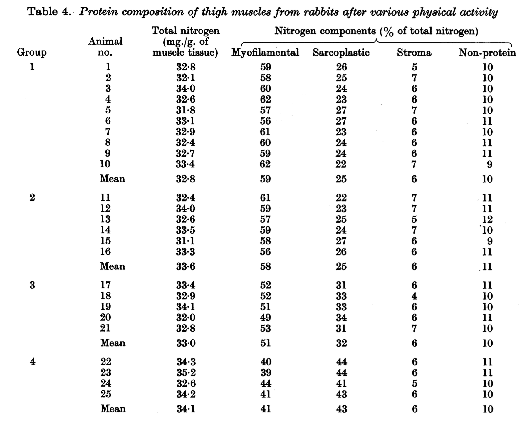
So at this point, the savvier readers are asking themselves: “If they could do this type of biochemical analysis back in the ’60s, how the heck don’t we have a clear answer about sarcoplasmic hypertrophy in humans yet in 2015?”
That’s a good question.
There are two main reasons, both of which are discussed in this paper.
- To do this type of biochemical analysis, you need a pretty hefty tissue sample. The smaller the sample, the more room there is for error. Improved lab techniques may have solved this problem in the past 50 years, but at least at the time this paper was published, they needed to take an entire gram of muscle tissue to run the analysis. One gram of muscle tissue is roughly one cubic centimeter. I don’t think you’ll find many human subjects willing to let a researcher rip that large of a tissue sample out of their muscles.
- Histological techniques (basically analyzing muscle under a microscope) won’t be able to accurately detect changes in sarcoplasmic versus myofibrillar protein densities. To quote the authors: “It would be feasible to carry out such an investigation with the aid of histological methods, but they have two major disadvantages. First, fixation and staining are accompanied by shrinkage of sections, rendering impossible sufficiently precise determinations of the proportions of sarcoplasm and of myofilaments in the total cell volume. Secondly, the myofilaments are forming myofibrils which are not clearly separated from the sarcoplasm in sections studied by histological methods. Electron microscopical examination has disclosed that myofibrils have no envelope; moreover, it is known that various chemical constituents of sarcoplasm, such as creatine phosphate, adenosine triphosphate and mitochondrial products, are capable of freely passing into and out of the myofibrils. Also, large molecules, for example inulin with a molecular weight of 6,000, can penetrate into the myofibril. It is difficult to tell whether the larger sarcoplasm-protein molecules actually pass into the myofibrils; but, at least theoretically, there is room for them between the myofilaments, which in the I bands are separated by wide spaces as judged by the lack of staining with osmium. Hence, methods other than histological seem to be preferable for studying the relationship between sarcoplasmic volume and myofilamental volume.”
So, keeping in mind that we probably aren’t going to get a tidy answer based on direct evidence, we need to turn to indirect evidence, which comes from two different sources: measuring intramuscular water concentrations, and examining functional characteristics of individual muscle fibers.
There are, to my knowledge, only two studies examining changes in intramuscular water after strength training.
The first one, published in 2014, did find an increase in intramuscular water concentrations, which may lead one to believe that sarcoplasmic hypertrophy had happened. However, I’m not sure that we can put a ton of stock in it.
The biggest issue with the study was simply that the participants didn’t gain very much muscle in the first place. The men gained about 1.3 kg. of muscle, and the women gained about 0.8 kg. of muscle. This was a 12-week study on untrained participants. In other words, the amount of muscle they gained was so small, especially considering the circumstances, that I just don’t think we can take much away from this study. Any amount of sarcoplasmic hypertrophy would necessarily be trivial because, well, the total amount of muscle they gained was trivial.
The second one is a classic from 1982. The researchers trained participants for six months and examined their muscle pre-training and post-training, and compared their muscles to those of elite powerlifters and bodybuilders. The powerlifters and bodybuilders were lumped together, so it’s impossible to draw a distinction between them, but that does give us a reference point for people who are very highly trained.
After six months of training, the participants gained a ton of muscle. In fact, the size of their individual muscle fibers had nearly caught up with the elite lifters’, so lack of growth isn’t a problem in this study.
The participants’ myofibrillar density slightly decreased and their sarcoplasmic volume relative to fiber size slightly increased over the course of the study. The changes were small, but reached statistical significance. When comparing the participants to the group of elite lifters, those trends were even more stark. The elite lifters’ myofibrillar density was way lower (almost 10% lower), and their sarcoplasmic volume relative to fiber size was way higher (about 10% higher). In other words, a tiny bit of sarcoplasmic hypertrophy occurred in the participants, and while you can’t infer a causal relationship since the elite lifters didn’t undergo an intervention as well, it certainly seems like quite a bit more sarcoplasmic hypertrophy separated the participants and the elite lifters.
Also, note that mitochondrial density decreased over the six months of training, and was even lower in the elite lifters. 6 of the 7 elite lifters also used or had used steroids. More on both of those things a bit later.
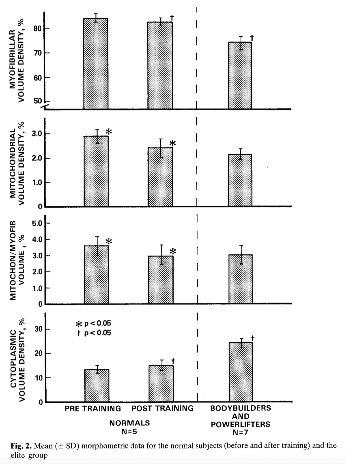
(You may be asking yourself, “How did they determine myofibrillar and sarcoplasmic volume if doing so requires such a large tissue sample?” I’m honestly not sure; it wasn’t clear in the methods section. They cite two other papers that apparently describe the method in more detail, but I don’t have access to them. Apparently they used electron microscopy, which doesn’t require as large of a tissue sample, so this may be more easily study-able these days.)
Now, let’s turn our attention to functional data.
You can estimate myofibrillar density by measuring the amount of force a single muscle fiber can produce, and dividing that force by the cross sectional area of the muscle fiber. This is called the specific tension of the fiber. A higher specific tension means higher myofibrillar density at face value, and a lower specific tension means lower myofibrillar density (and thus a higher proportion of sarcoplasm), in most circumstances. Typically, the only major factor that can throw off this relationship in non-fatigued muscle is post-translational modifications of contractile proteins. In other words, if the actin and myosin that cause muscle contraction are modified in some way that causes them to not function properly, that can cause a drop in specific tension independent of myofibrillar density. But, in most circumstances (because post-translational modification is pretty rare), specific tension gives you a pretty good estimate of myofibrillar density.
Now, you may balk at the idea of trying to draw any conclusions from studies of single muscle fibers, but bear in mind that using single fibers is one of the only ways to mitigate the effects of a whole host of confounding factors, including (to quote a study on this very topic), “differences in intramuscular fibre orientation or pennation, the presence of intramuscular connective tissue, differences in mechanical leverage related to joint position, the possible coactivation of antagonist muscles during strength testing, and variation in motor unit recruitment patterns, central drive and subject motivation.”
A recent study compared individual muscle fibers from bodybuilders, power athletes (American football players, track and field athletes, and weightlifters), and control subjects.
The bodybuilders had the largest muscle fibers by a mile (88% larger than the controls, and 67% larger than the power athletes). The bodybuilders’ muscle fibers produced more force than the control subjects’, but they actually produced slightly less force than the power athletes’ muscle fibers (the difference in total force between bodybuilders’ and power athletes’ fibers was not statistically significant, however).
Here was the really interesting part: Per unit of cross-sectional area, bodybuilders’ muscle fibers produced way less force than either the power athletes’ or the controls’ (66% less than the power athletes’, and 41% less than the controls’).
After obtaining this result, the researchers performed another analysis to see whether post-translational modifications explained the difference, and concluded any possible post-translational modifications played, at most, a minor difference.
In other words, the bros were right all along. Training like a bodybuilder causes nonfunctional sarcoplasmic hypertrophy.
Right?
Not so fast. There are three problems with jumping to that conclusion from this study:
- There wasn’t actually an intervention. It could just be that people with a particular type of abnormal muscle fibers are more likely to become bodybuilders.
- The sample sizes for this study were pretty small.
- This little line is telling: “A negative trend between fiber cross-sectional area and specific tension has been reported before in single muscle fibre segments of untrained people and frogs. In the present study, this negative trend is apparent across all groups (emphasis mine). It has been suggested that this may be related to accumulation of inorganic phosphate, due to longer diffusion times from the interior of the fibre to the surrounding incubation medium.”
The third point is probably the most important point.
While total muscle cross-sectional area generally correlates quite strongly with capacity for torque production, single fiber studies tell a different story.
This one in particular illustrates the difference. In the largest study on single fibers that I’m aware of, the researchers actually found that capacity for force production was more strongly related to muscle fiber diameter than cross-sectional area. They plotted peak force against diameter on a log/log scale, and found that the slope for both type 1 and type 2 fibers was very close to 1. As you’ll remember from a previous article, the slope of a log/log graph tells you the exponential relationship between two variables. In the previous article on allometric scaling, the slope of the line comparing mass versus strength was 2/3 and the slope of the line comparing mass versus metabolic rate was 3/4. If the slope is 1, that means the relationship is linear, not exponential (since raising something to the first power doesn’t do anything).
Most importantly, they concluded that the slope of the line of best fit for that log/log plot was almost certainly not 2, even when accounting for variability and potential error. If the slope was 2, that would mean that peak force scaled with the square of the diameter, which, in turn, would mean that peak force scaled linearly with cross-sectional area. In other words, since there’s almost no way that the slope of the log/log plot was 2 (p<.0001 for both type 1 and type 2 fibers), that means you should expect the peak force of a single fiber to be more closely related to fiber diameter than cross-sectional area.
Through that lens, let’s look at the data from the study comparing bodybuilders, power athletes, and controls again.
Since absolute values weren’t reported for all measures, I made both cross-sectional area and peak force relative to the controls, and just used arbitrary units. I maintained the relationships (percentage differences) reported in the study.
| Controls | Power Athletes | Bodybuilders | |
| Cross-sectional area | 10 | 11.26 | 18.8 |
| Diameter (calculated) | 3.57 | 3.79 | 4.89 |
| Peak Force | 1 | 1.5 | 1.33 |
| Predicted Peak Force | 1 | 1.06 | 1.37 |
Using the more appropriate relationship (force relative to fiber diameter, instead of cross-sectional area), we see a different picture from the one painted above. The bodybuilders’ muscle fibers produced almost the exact same amount of force per unit of diameter as the controls’ fibers (bottom row – the bodybuilders’ fibers only produced about 3% less force per unit of diameter than the controls’). This is in contrast to the 41% difference when using specific tension (force relative to CSA). The power athletes’ fibers still had way higher peak forces relative to diameter than either of the other groups.
In other words, the issue wasn’t that the bodybuilders were doing something “wrong” that decreased force relative to CSA; based on how much larger their muscle fibers were than controls’, their muscles produced about as much force as you’d expect based on fiber diameter. It’s more likely that either the power athletes were doing something “right” to increase force relative to fiber dimension above what would be expected, or that people whose fibers are naturally capable of producing more force than they “should” are more drawn to being power athletes in the first place – likely a combination of both factors. But keep in mind, even for power athletes, specific tension decreased as fiber size increased.
However, this leads us to another quandary. Since myofibrillar density should still scale linearly with cross-sectional area, but muscle force scales linearly with diameter instead … does sarcoplasmic hypertrophy simply go hand in hand with hypertrophy itself? Is an increase in or maintenance of myofibrillar density actually the “weird” thing to happen?
Maybe.
But since I doubt we’ll have many more single-fiber studies in the near future, because of how time and labor-intensive they are, and how low a priority this type of research is for serious athletes. It would be directly relevant to geriatrics though (so if there are any exercise physiologists reading this who work with elderly people, consider this a subtle hint). For the time being, though, the best we can do is look at whole muscle studies, and muse about mechanisms.
The first study that comes to mind is this one on elite level powerlifters that found very strong correlations between muscle thickness (rather than cross-sectional area) and strength.
Most studies show that strength trained people’s whole muscles have higher specific tensions than those of untrained people. However, this study (to the best of my knowledge) is the only one that compared whole muscle specific tension (force/CSA) to single fiber specific tension pre- and post-training. They found that specific tension for single fibers was unchanged, but specific tension for the whole muscle increased. They posited that lateral force transmission (muscle fibers side-by-side linking up with each other to aid in force transmission) was the most likely cause for the increase in whole-muscle specific tension, and that myofibrillar density within the individual fibers themselves was unchanged since single fiber specific tension didn’t change.
This study and this study had similar results for single fibers (unchanged specific tension) pre- and post-training, but the researchers didn’t compare those results to changes in whole-muscle specific tension. This study and this study on the other hand, observed an increase in whole-muscle specific tension and single fiber specific tension without any overall muscle hypertrophy, although the second study was confounded by the fact that it was performed on elderly subjects, who naturally experience a decrease in specific tension with age (so the increase in specific tension just got them back to “normal”).
There are also a host of other factors, such as increased muscle activation, decreased activation of antagonist muscles, and even changes in muscle architecture that favorably alter the muscle moment arm that could increase force production relative to CSA as well.
With those confounding factors in mind, high-intensity training does seem to produce larger increases in specific tension than lighter training does, and bodybuilders regularly produce less force per unit of muscle CSA than strength athletes do, and sometimes even less than untrained controls. In this study as well, there was a negative correlation between CSA and specific tension. In this study, on the other hand, bodybuilders actually produced more knee extension torque per unit of CSA than powerlifters did.
On the whole, it seems that strength training increases force/CSA for the whole muscle, but likely doesn’t affect force/CSA (and myofibrillar density, by extension) at the level of individual fibers unless no actual muscle growth takes place. On the other hand, in most studies, bodybuilders’ force/CSA is lower than strength athletes’, and is often similar to untrained controls. This is in line with the broader observation that force relative to size is most strongly related to fiber diameter, and that it generally decreases as fiber size increases. Strength training bucks that trend and maintains the relationship between force and CSA, but bodybuilding-style training simply seems to maintain the trend of specific tension decreasing with increasing fiber size, as seen in larger studies.
So, does this mean that sarcoplasmic hypertrophy (an increased percentage of sarcoplasmic proteins relative to myofibrillar proteins) typically goes hand in hand with muscle hypertrophy?
Possibly.
Remember, there are still a few other potential confounding factors. Most notably, the fact that larger fibers may accumulate inorganic phosphate. Inorganic phosphate directly decreases the force of muscle contraction by screwing with how well myosin can bind to actin, so that would account for a decrease in force/CSA without a change in myofibrillar density. However, keep in mind that this is just a proposed mechanism, not a proven one in this particular context. Usually inorganic phosphate only accumulates as the muscles fatigue (when you’re splitting phosphates off ATPs faster than your energy systems can replenish ATP levels); however, it’s known that inorganic phosphate accumulation is one of the main reasons force/CSA decreases after limb immobilization, so it’s possible for it to accumulate in non-fatigued fibers under some circumstances.
There is a potential rationale for levels of inorganic phosphate rising in the muscles of bodybuilders – inorganic phosphate can act as a buffer when muscular pH drops during training. Maybe bodybuilders’ muscles accumulate inorganic phosphate as a response to high rep training that causes local muscular acidosis. However, it’s one of the less important cellular buffers, and even if that would explain how inorganic phosphate accumulated in bodybuilders’ muscles, that still wouldn’t help explain the more general trend of decreased force/CSA with increasing fiber size, especially in type I muscle fibers (which rely more on aerobic metabolism, and don’t reach as low of pHs).
Let’s just assume for the moment that accumulation of inorganic phosphate plays, at most, a minor role. Let’s also assume that post-translational modifications don’t play a major role (and they generally don’t, outside of elderly populations and animals that don’t produce myostatin). The only other option (that I’m aware of) to explain the declining force/CSA with increasing fiber size is a decrease in myofibrillar density. In other words: sarcoplasmic hypertrophy.
So now we need to ask ourselves: “Why would this happen in the first place? And why might it happen more with bodybuilders than strength or power athletes?”
The simplest explanation I can think of: energy.
A hefty portion of the sarcoplasmic proteins are involved in the various steps of anaerobic metabolism. As a muscle fiber gets larger, it naturally relies less and less on aerobic metabolism for two major reasons:
- Unless you’re doing dedicated aerobic training, mitochondrial density (mitochondria are the sites of aerobic metabolism) generally decreases. This was seen in MacDougall’s study; mitochondrial density dropped over six months as the participants’ muscle grew, and it was lower yet for the elite lifters.
- The ratio of capillaries per unit of muscle fiber CSA generally decreases with increasing fiber size, unless you’re doing dedicated aerobic training. This means you can’t get as much oxygen to the mitochondria closest to the myofibrils further inside the muscle fiber since there’s a longer diffusion distance from the muscle cell membrane (the sarcolemma) to the mitochondria deep inside the cell. This, in turn, favors a shift toward anaerobic metabolism.
This idea makes sense in light of the single fiber study from earlier comparing bodybuilders, power athletes, and controls; not only were the bodybuilders’ fibers much larger, but they were also the only group not doing dedicated cardiovascular training.
However, it doesn’t fully explain why specific tension (and thus myofibrillar density) wouldn’t drop off with heavy strength training, but may with lighter, higher volume bodybuilding-style training. Increased training volume in general, and particularly increased training close to failure with lighter loads imposes greater energetic demands on the muscle, which should help spur on some of those aerobic adaptations (increased capillary density and increased mitochondrial density) that would decrease the need for increased levels of proteins associated with anaerobic metabolism.
I think (though I realize that this explanation is flimsy) that since bodybuilding-style training is both more aerobically and anaerobically taxing, the hugely increased anaerobic demands may lead to larger anaerobic adaptations that outweigh the increased aerobic adaptations. With heavy triples, you’re barely going to burn through your stored ATP and phosphocreatine, so you don’t need to get a ton of your energy of anaerobic glycolytic metabolism; that’s entirely different when you’re hitting multiple challenging sets of 8-15 reps.
Looking at changes in sarcoplasmic protein metabolism after different workouts and specifically separating mitochondrial proteins from other sarcoplasmic proteins may help shed some light on this idea, but some studies include mitochondrial proteins in the sarcoplasmic protein fraction (like this one, which showed that sarcoplasmic muscle protein synthesis was elevated 24 hours post-workout after training to failure with 30% 1rm loads, but not 90% 1rm loads, or this one, which showed that a slower lifting cadence caused larger increases in sarcoplasmic protein synthesis than faster lifting cadences), and others separate mitochondrial proteins out and only analyze the non-mitochondrial sarcoplasmic proteins (like his one, or this one, which showed that a slower lifting cadence caused larger increases in sarcoplasmic protein synthesis than faster lifting cadences).
One last thing before wrapping up: I know I’m going to get a question or 30 about steroids. Do they contribute to sarcoplasmic hypertrophy?
Bro wisdom says yes. A lot of people report gaining a lot of weight very quickly when they go on steroids (much more than could be explained by plain old myofibrillar hypertrophy) and losing it very quickly when they come off, likely indicating shifts in the amount of water they’re storing. However, it’s not always apparent that the water is being stored in muscles themselves (some people report feeling like their muscles are “full,” while other perceive it to be more like bloating), and thus far, the two studies that have actually looked into this issue reported no increases in intra-muscular water (one on testosterone, and one on nandrolone).
However, dosing may be an issue: The dosages people run in the real world rarely get studied, for ethical reasons. Also, in MacDougall’s study, sarcoplasmic volume relative to fiber volume was much higher for the elite lifters (most of whom used steroids) relative to the normal participants in the study. On the other hand, in the single fiber study comparing bodybuilders to power athletes and untrained controls, they didn’t find any significant differences between the fibers of the bodybuilders who reported steroid use and those who didn’t report steroid use. However, in this study, drug-free strength athletes could produce almost 50% more force per unit of quad mass than bodybuilders on steroids could (though training differences likely factor in as well).
In other words, it’s hard to say for sure what role, if any, steroids play.
So, just to wrap this sucker up:
- Sarcoplasmic hypertrophy has never been directly, convincingly measured in humans – at least not up to the standards it would take to convince me personally. One study showed increases in intracellular water post-training, but the overall hypertrophy was very small in the study, so I’m not ready to put too much faith in it. Another study did show a very small degree of sarcoplasmic hypertrophy along with very robust overall hypertrophy after six months of training, but due to the very tiny degree of sarcoplasmic hypertrophy measured, it’s also probably not worth putting a whole ton of faith in. The elite lifters in that study did have way more sarcoplasmic volume relative to muscle volume, but since they weren’t involved in an intervention, it’s impossible to know whether that difference was due to training, or due to pre-existing differences.
- Though non-protein solutes in the muscle (like glycogen) could potentially contribute slightly, any effect they could have would be small and transient. An increase in the proportion of sarcoplasmic protein relative to myofibrillar protein would be the much more likely cause, if sarcoplasmic hypertrophy did occur.
- We know that sarcoplasmic protein synthesis and breakdown aren’t directly hitched to myofibrillar protein synthesis and breakdown, and that in mammalian muscle, while a 3:1 myofibrillar:sarcoplasmic protein ratio is typical, a 1:1 ratio has been observed (albeit, in middle aged rabbits) without any obvious ill effects.
- Peak force of single muscle fibers is more closely related to muscle fiber diameter than muscle fiber cross-sectional area. In general, as CSA increases, force relative to CSA decreases. Without alternative explanations (all of which are unlikely under most circumstances in healthy, young people – things like inorganic phosphate accumulation and post-translational modifications of contractile proteins), the most obvious reason for this decrease is a decrease in myofibrillar density – sarcoplasmic hypertrophy.
- Though the picture painted by the current scientific literature is still hazy, it seems that strength training preserves the relationship between force and CSA as the fibers grow, but bodybuilding-style training may not. However, some of the studies that showed increased force/CSA used lighter loads (60%1rm), and one study even showed bodybuilders’ quad torque/CSA was higher than powerlifters’.
- It could just be that the odds of sarcoplasmic hypertrophy increase as muscle fibers get increasingly larger. As fibers grow, capillary density relative to fiber area decreases, and mitochondrial density decreases, increasing the reliance on anaerobic metabolism, which is carried out by sarcoplasmic proteins. Even in elite powerlifters, it was muscle thicknesses that correlated very tightly with strength (although that study didn’t measure CSA as well – it could have revealed just as strong of correlations).
- Based on the current evidence on highly trained subjects, there are too many confounding factors to tell whether different training styles increase the odds of sarcoplasmic hypertrophy. In vivo measurements are influenced by muscle attachment points, muscle activation, co-contraction of antagonist muscles, motivation, muscle moment arms, pennation angles, etc. Thus far, most of the direct comparisons we have are in vivo comparisons; so far there aren’t many single-fiber studies to tease out what’s going on at the cellular level (which is where you’d need to look if you wanted to study sarcoplasmic hypertrophy). The only study we have comparing the muscle fibers of bodybuilders and power athletes seems to indicate sarcoplasmic hypertrophy in the bodybuilders, but that could be due to innate differences independent of training, or simply be related to how much larger the bodybuilders’ muscle fibers were.
I’ll admit – I’m finishing this article with an opinion much different from the one I started with. As of about 5 days ago, I was confident that sarcoplasmic hypertrophy was either a myth or, at most, played only a trivial role in overall muscle hypertrophy. Now I’m confident that it happens and, while I’m not totally convinced, I’m much more open to the idea of it having a notable effect on overall muscle growth. I’m still skeptical that training in a specific manner will either minimize the “risk” of sarcoplasmic hypertrophy (from the perspective of a powerlifter) or increase your chances of sarcoplasmic hypertrophy (from the perspective of a bodybuilder), but I’m open to the idea that it could.
Ultimately, this is a question that won’t be settled until we have a lot more high-quality single fiber studies (or unless some brave souls want to donate a cubic centimeter of their muscles to science). People with strong backgrounds in muscle physiology come down on both sides of this debate. Dr. Stuart Phillips and Dr. Anders Nedergaard think it’s hogwash, Dr. Brad Schoenfeld thinks it happens, but that it plays only a small role in overall muscle growth, and Lyle McDonald seems convinced that it happens and that it can play a meaningful role in muscle growth.
Hopefully, at the very least, this article gave you a deeper understanding of the subject and raised some questions you hadn’t thought to ask.
If you liked it, be sure to share it with your friends who have a die-hard position on this subject, whichever way they lean.
Addendum, June 2017
A 2007 study by Cribb et al adds another wrinkle to this discussion. I missed it when looking for studies initially because it’s more of a supplement study (looking at the effects of creatine and whey) than a muscle physiology study.
The participants were recreational bodybuilders who clearly had a decent amount of training experience; the average bench press at the beginning of the study was ~100kg/225lbs. They were split into four groups. One group was supplemented with carbohydrate, one group was supplemented with carbohydrate and creatine, one group was supplemented with whey protein, and one group was supplemented with whey and creatine. They trained using the Max-OT program for 11 weeks (as an aside, the use of Max-OT as their training program made me chuckle a bit. I don’t see as many people recommending it nowadays, but it was a really popular style of training when this study was conducted. It would be like seeing a new study using 5/3/1 as the training program).
The study examined (among other things) contractile protein content: the amount of contractile protein per unit of mass. If contractile protein content increased, that would be evidence of relative myofibrillar hypertrophy, while if it decreased, that would be evidence of relative sarcoplasmic hypertrophy.
The groups taking whey and/or creatine all had larger increases in contractile protein content than the group just taking a carbohydrate supplement (p<0.05). Furthermore, the two groups taking creatine tended to increase contractile protein content more than the group just taking whey (p=0.07-0.08).
There are two other interesting things to note from this study:
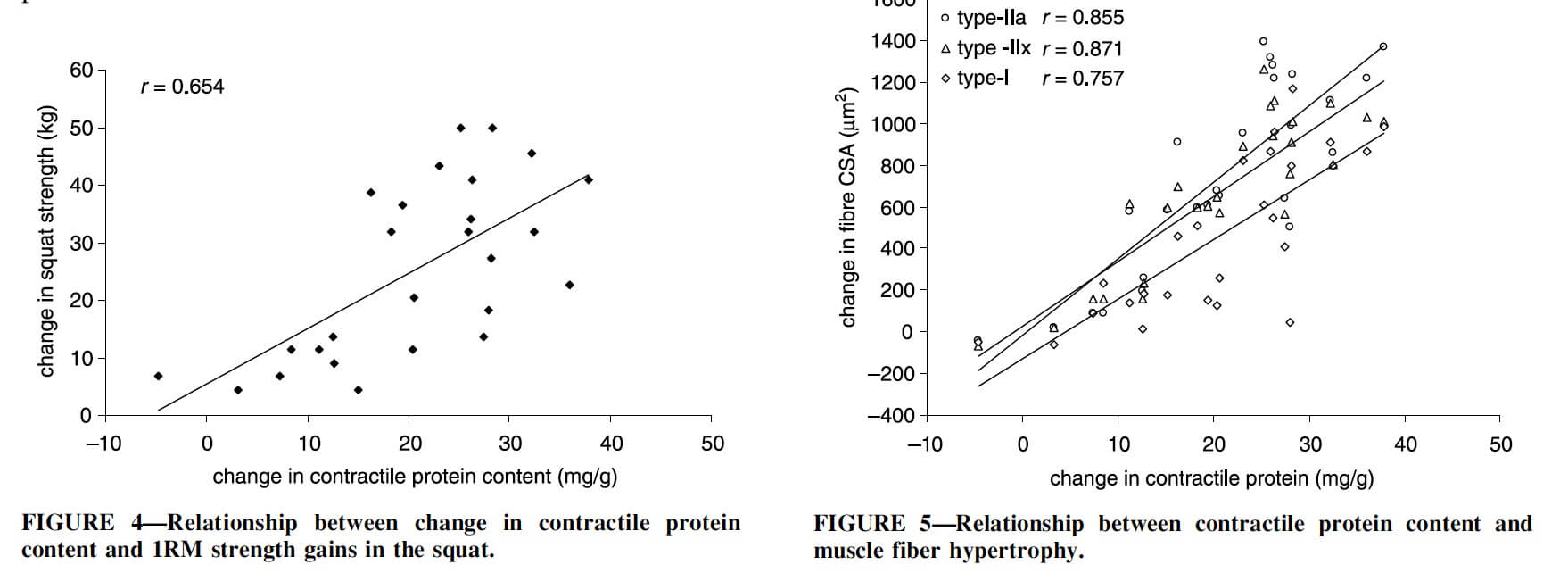
Increases in contractile protein content were independently associated with increases in squat 1RM, and increases in contractile protein content were strongly correlated with hypertrophy of all three major muscle fiber types. That means that in this study, myofibrillar hypertrophy was the primary driver of general fiber hypertrophy.
However, it’s also worth noting that the changes in contractile protein content were quite small: up to 40mg/g. That means the proportion of muscle fiber weight accounted for by contractile proteins only increased by 4% at most. Though that’s a relatively small increase, it still stands in contrast to the MacDougall study, which found a slight decrease in contractile protein content with training. This may give us evidence that training style actually affects the ratio of sarcoplasmic and myofibrillar hypertrophy. The subjects in the Cribb study trained with sets of 4-6 reps, while the subjects in the MacDougall study trained with sets of 8-10 reps. It’s always tenuous to compare between studies, though, so certainly don’t take this as proof that training style affects sarcoplasmic vs. myofibrillar hypertrophy. Furthermore, a study by Moore et al found that while protein consumptions affects both myofibrillar and sarcoplasmic protein synthesis, strength training (10 sets with 8-10rm loads) only affects myofibrillar protein synthesis. However, the question of training style remains: the sarcoplasmic protein synthesis response may have been different with a different training intervention.
Finally (and unrelatedly), this is a good study to share with people who claim that creatine only causes muscle growth via an increase in water content in the muscle (i.e. sarcoplasmic hypertrophy). The groups taking creatine in this study had the largest increase in contractile protein content; even if water content increased, that increase was outpaced by the observed increases in contractile protein content.
Addendum, January 2018
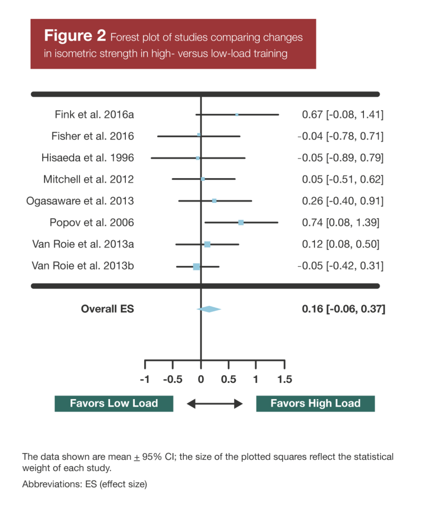
A recent meta-analysis by Schoenfeld et al looking at the effects of training load on hypertrophy, dynamic strength, and isometric strength helps counter one of the main arguments people use to contend that light, high rep training causes sarcoplasmic hypertrophy. People claim that since strength gains are larger with heavier training, heavy training must be adding more contractile proteins (myofibrillar hypertrophy), while lighter training must be expanding muscle size without adding as many contractile proteins (sarcoplasmic hypertrophy). Earlier in this article I discussed why that’s not an entirely logical argument, but this meta-analysis provides us with some direct evidence to refute it.
Unsurprisingly, heavy training was better for dynamic strength. However, there’s a skill component to dynamic strength, and heavier training helps to train that skill. On the other hand, there was no significant difference between high load and low load training for gains in isometric strength (i.e. force output with virtually no skill required). This suggests that low load training is still adding contractile proteins just as effectively as high load training; it’s just not great for training you to use them effectively for maximal dynamic contractions (i.e. 1RMs).
This meta-analysis was discussed in more detail in Volume 1, Issue 7 of MASS.
